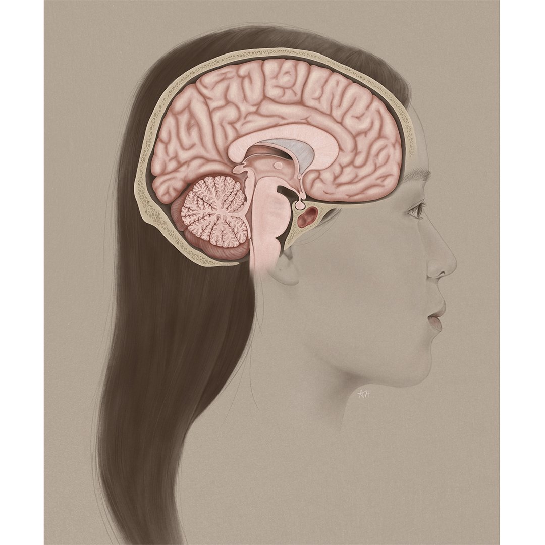
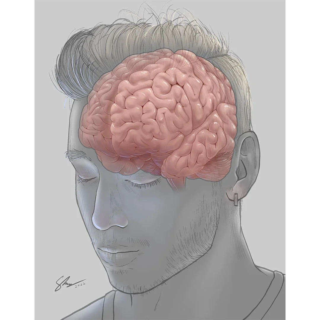
"When Neuro was a first-year course, the first assignment was just a very straightforward illustration of the brain in isolation," says Shelley Wall, associate professor at the University of Toronto.
Until this year, students in the Master of Science in Biomedical Communications program created original but standard illustrations of brains, in whole or in section. The illustrations consolidated their knowledge of the anatomy and developed their digital rendering skills.
Rescheduling the course Neuroanatomy for Visual Communication from first-year curriculum to second-year gave Wall, the course instructor, the opportunity to challenge the design and rendering skills of the graduate students.
Innovating course content
"In first year, students are still finding their way in anatomical illustration. Now that Neuro is a second-year graduate course, I could take it to the next level because the students' skills are so much more developed. So I conceived the neuro self-portrait assignment," she says.
In Neuroanatomy, students learn the structure and function of the brain and the cranial nerves. They engage in interactive exercises, examine brain specimens and skulls, study historical and contemporary texts, and watch videos of dissections.
"Drawing the brain is one thing. You must make it accurate. But what makes this assignment so different is that you really must understand all the important relationships between the brain, the brain case, and the external features of the head. And making the assignment a self-portrait is a way of making it also a completely unique illustration that really puts the students’ stamp on it and allows them some creativity," she says.
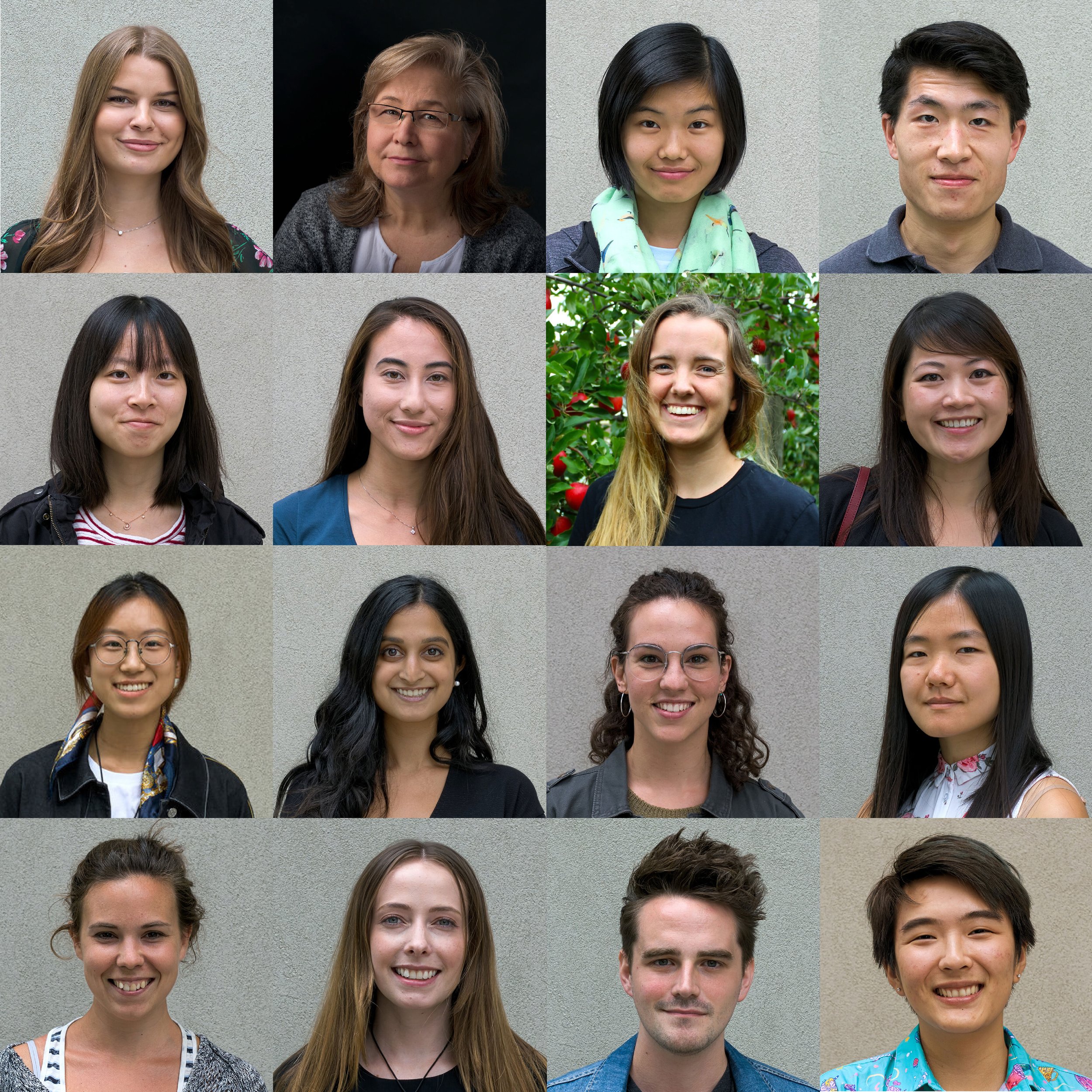
Exceeding expectations
Even working within the constraints of the course assignment, and the strict parameters of depicting the brain with accuracy, the second-year graduate students delivered a broad range of unique and original illustrations.