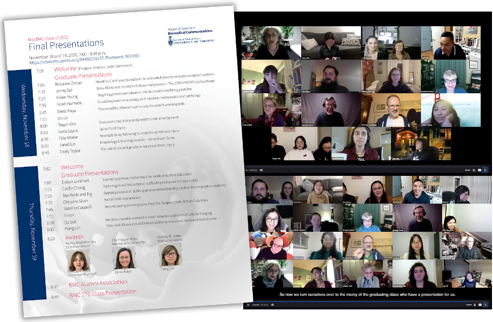
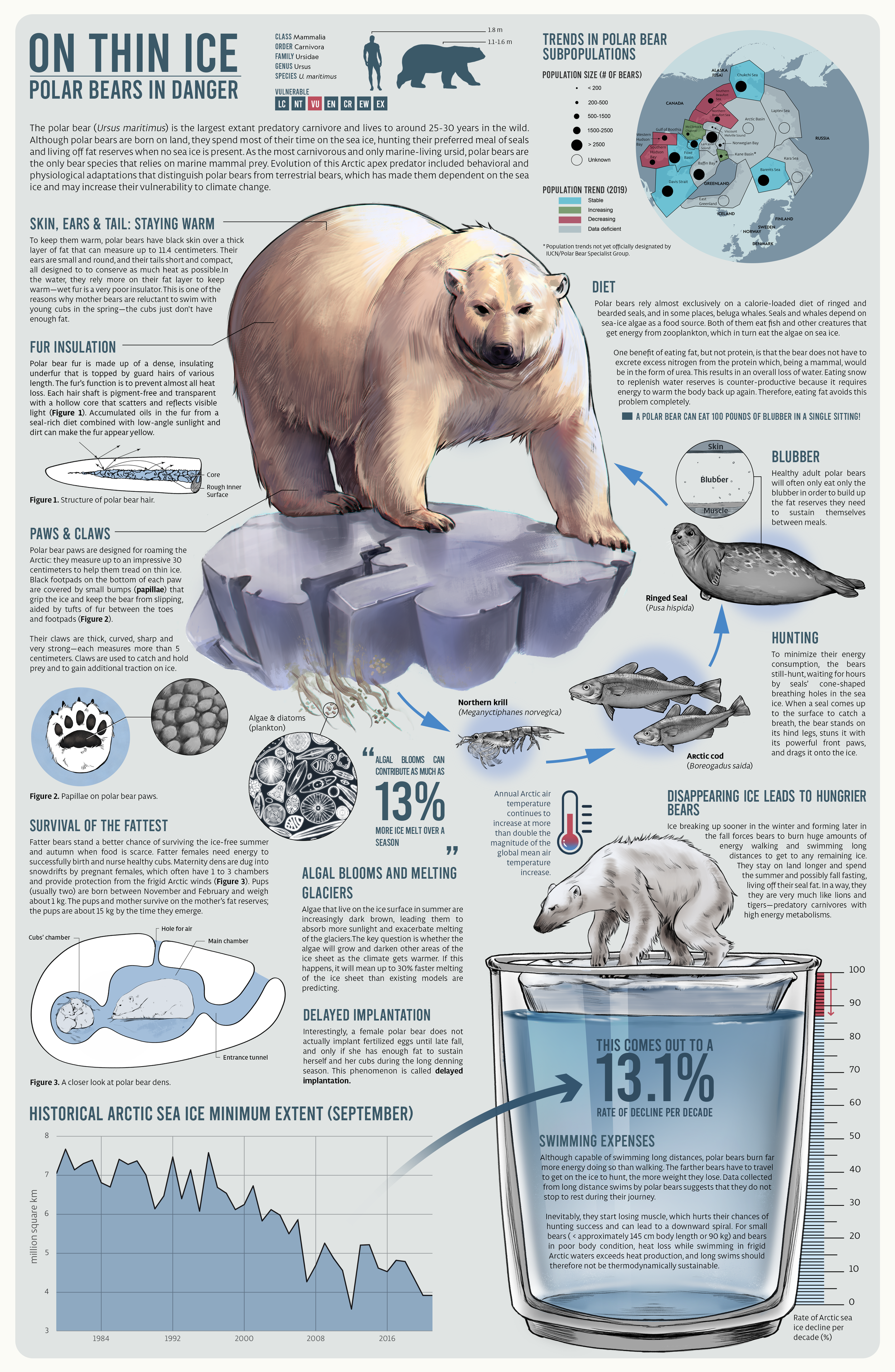
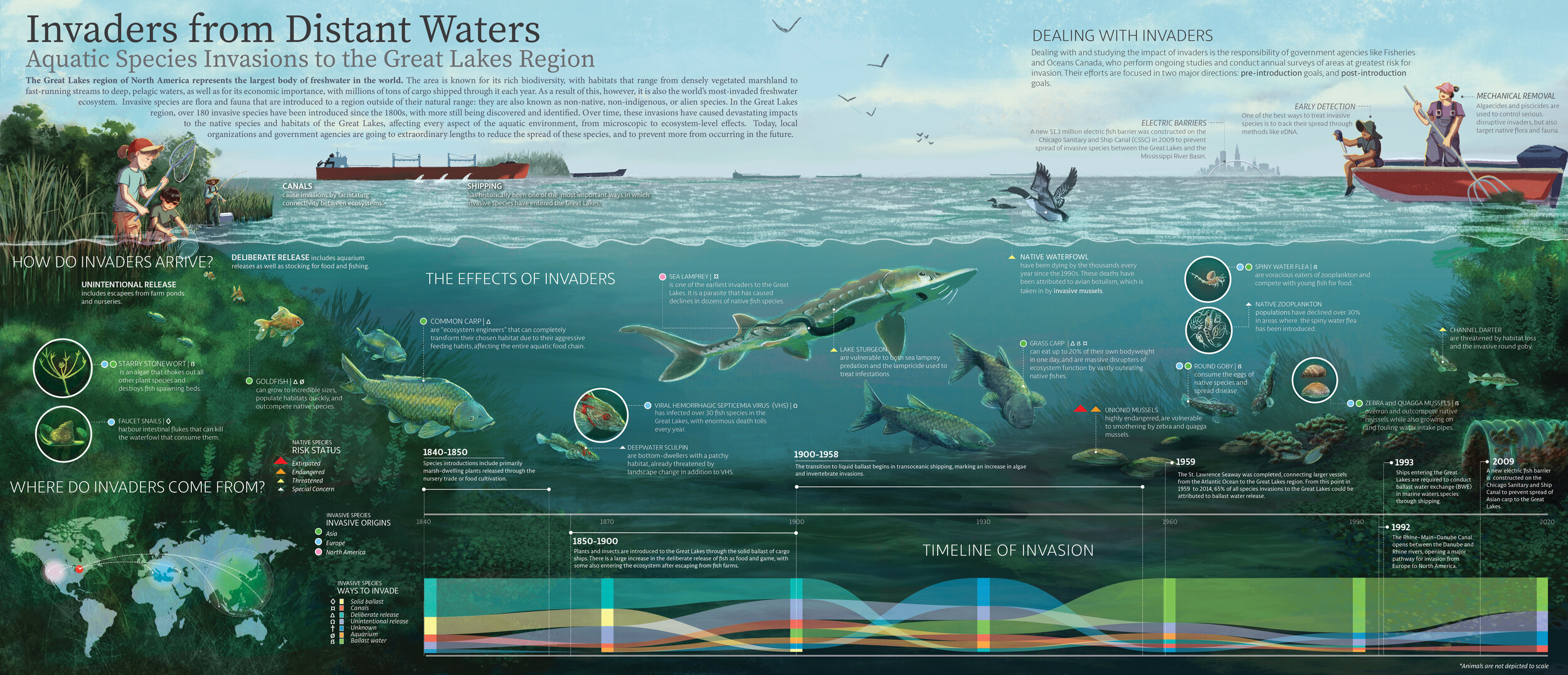
Printing skull models for a virtual classroom

12:00 p.m.

Maeve Doyle

Professor Dave Mazierski spent the Christmas 2020 break at home printing 3D-models of bat-eared fox skulls. He made them for each of the nearly 50 undergraduate students attending his scientific drawing class online.

3D-prints of a bat-eared fox skull. Photo credit: D. Mazierski

3D-prints of a bat-eared fox skull. Photo credit: D. Mazierski

Mazierski, a vertebrate palaeontologist and associate professor of biomedical communications at the University of Toronto, teaches HSC302 Biocommunication Visualization. Completing an accurate drawing of a skull is one of the course’s exercises.

Normally, a biology technician provides primate skulls for the students to observe and sketch. Mazierski would provide transparent grids through which to observe the models, clips to support the grid, and drawing materials. Rows of drawing stations would be set up in one of the biology department’s laboratories.

“Like a skull-sketching factory,” he says.

Pre-pandemic skull-sketching laboratory. Photo credit: D. Mazierski

Pre-pandemic skull-sketching laboratory. Photo credit: D. Mazierski

But with teaching forced online by the global COVID-19 pandemic, he needed a substitute for this learning experience. There wasn’t a space large enough to accommodate physical distancing. The real primate skulls were too valuable and too fragile to lend.

Mazierski got approval from the director of the Master of Science in Biomedical Communications to borrow the program’s Form2 3D printer. He researched online repositories for 3D data for skulls, and settled on the bat-eared fox.

“It’s got interesting teeth.” He holds up a resin model of the skull. “It’s slightly smaller than life, but it’s large enough that students can see the features we want them to understand and to draw the various elements of the skull. The size and shape was easy to print as a single object and to clean.” He taps the desk with the 3D print. “And they’re durable,” he says.

Mazierski printed the models in his newly-converted 3D-printing studio, formerly his daughter’s bedroom.

“It took 22 hours to print them nine at a time–” the maximum the desktop 3D-printer could produce ”–and another 20 minutes per skull to remove them from the build platform and clean them,” says Mazierski.

Professor Dave Mazierski separates a 3D print of a bat-eared fox skull from the build platform. Photo credit: D. Mazierski

Professor Dave Mazierski separates a 3D print of a bat-eared fox skull from the build platform. Photo credit: D. Mazierski

He then faced the problem of getting the skulls to students dispersed by the pandemic. He packed the models into envelopes, along with grids and a stand. He also fabricated the stands himself from pine boards.

Students collected the majority of the envelopes from lockers in the biology department, but some supplies had to be mailed to students attending class from such places as British Columbia, Ireland and Pakistan.

Drawing to learn in science

Laboratory drawings are a standard part of life science studies. In comparative anatomy courses, for example, students are expected to create manuals in which they draw their observations. “In vertebrate palaeontology, you have to be able to draw your specimen to report it for publication,” he says.

Encouraging people to draw is sometimes a way to encourage them to learn in a different way. “While you’ve got to draw the line somewhere, a scientific drawing can’t be vague or ambivalent. If it’s going to be accurate and convey information, all those lines must have meaning,” he says. “The act of drawing forces you to understand.”

BMC prof receives IMS Course Lecturer Award

1:10 p.m.

Michael Corrin, associate professor teaching stream, Biomedical Communications, has been recognized again by the Institute of Medical Science (IMS) at the University of Toronto for excellence in teaching.

Corrin has been named the 2020 recipient of the IMS Course Lecturer Award. The award is presented annually for sustained contribution of three years or more to excellence in lecturing in an IMS graduate course.

MScBMC Associate Professor Michael Corrin smiling at the camera.

Corrin is also the 2017 recipient of the IMS Course Director Award for sustained excellence in the teaching, administration and development of a IMS graduate course.

On behalf of his colleagues and students, past and present, the Biomedical Communications program thanks Professor Corrin for his many contributions and dedication to training highly qualified personnel in science communication.

Class of 2020 joins the BMC family of alumni

8:14 a.m.

The Master of Science in Biomedical Communications welcomes the newest cohort of graduands to their family of BMC alumni today.

The Class of 2020 made final presentations of their Master's Research Projects (MRPs), online and over two evenings, November 18 and 19.

A presentation agenda layered above screen captures of MScBMC Class of 2020 students in a video call over Zoom..

MRPs ranged from 3D animations of public health issues to interactive e-learning for undergraduate education to graphic narratives for patient education.

The final presentations are available to view here: 

November 18: https://youtu.be/-ODSpqU8Xh0
November 19: https://youtu.be/pXXHGnPpD2U

The Class of 2020 convocates Saturday, November 21 at noon. Watch the University of Toronto's Fall Convocation 2020 and Virtual Ceremony here: https://www.utoronto.ca/convocation.

Congratulations to the MScBMC Class of 2020 who graduate in the 75th year of the Biomedical Communications program!


Information design: more than pretty pictures

9:35 a.m.

Maeve Doyle

Year I students in the Master of Science in Biomedical Communications recently created information graphics in partial fulfilment of the course requirements for MSC2023H Information Design.

The graduate students' projects attempt to clarify complex medical or scientific subject matter for a specific target audience.

"Part of the whole process is understanding how to appropriately encode visual variables, and how to make information pop by manipulating pre-attentive features in the design of the graphic," says Jodie Jenkinson, course instructor, associate professor and director of the Biomedical Communications program.

In their first year, Biomedical Communications students are trained in several subjects including information design. They are taught how to create clear information hierarchies in the design of visual displays. This includes the purposeful ordering and integration of graphical elements and text.

Leveraging human visual perception

"One important goal of information design is to attract the attention of the viewer through the manipulation of visual elements. This increases engagement and furthers the impact of the communication,” says Jenkinson. "But more importantly, things that might be difficult to discern in a block of raw data can become crystal clear when you map that data to a graphical representations."

She says that human visual perception is attuned to pattern-finding and comparison. Trends and relationships can often be understood from a properly designed image far more easily than they can from a table of data or a paragraph of text.

The brain’s visual cortex, and the areas that surround it, have evolved to find patterns and make comparisons. “Biomedical communications specialists use information design to engage that uniquely powerful part of the brain.”

Information graphics produced by: Amy Assabgui (Ecosystem Architects), Viktoriya Khymych (On Thin Ice), Naomi Robson (The Plight of the Pangolin), Shay Saharan (The Most Endangered Great Ape–Orangutan), and Aimy Wang (Invaders from Distant Waters).

NSERC funds training in visual education of researchers

11:09 a.m.

Maeve Doyle

NSERC awarded Nicholas Woolridge, associate professor in the Master of Science in Biomedical Communications at the University of Toronto, a $20,000 grant in its pilot Science Communications Skills competition.

The grant will support Woolridge’s development of a workshop to train STEM students and researchers in the effective communication of science to the public. The workshop will be developed for, and with, principal investigators and their trainees.

Nicholas Woolridge, Associate Director, Master of Science in Biomedical Communications. Photo credit: Maeve Doyle

Nicholas Woolridge, Associate Director, Master of Science in Biomedical Communications. Photo credit: Maeve Doyle

DiSCOVER–Design in Science Communication: Opportunities in Visual Education for Researchers

Woolridge says that PIs, postdoctoral fellows and graduate students are often called upon to communicate scientific concepts. But, they may lack training in the skills they need to effectively communicate that science in visual form, such as data visualizations, imaging, graphs, diagrams, flow charts, and other forms of visual media.

“The primary goal of DiSCOVER is to design a prototype workshop experience to impart audience-aware design methods and skills,” he says. “We mean to create a portable workshop format that can be deployed beyond U of T for researchers and trainees in STEM.”

Design Process

Unique to DiSCOVER is the co-design process, which will involve the stakeholders. "By building a facilitated collaborative phase into the workshop development, we hope to address both the needs of the people we are training, and the needs of the audiences who will be informed by them." The project will partner with Bridgeable Inc., a Toronto design consultancy, on the co-creation training and process.

The benefits of the project are two-fold. "Participants will leave with greater experience in the design of visual media for science communication, but another takeaway will be the design of the workshop itself."

Portable Design

Woolridge will document the activities and exercises so that DiSCOVER can be implemented elsewhere. "It won't just be a one-time activity. If people have a good experience with it, facilitators in other research centres can mount the science communications training again and again, year after year."

Future funding could support work to refine the design for different domains. "The communications needs and objectives of, say, biology might vary between biological disciplines. Similarly, the needs and objectives will vary for different  domains, such as earth sciences or physics or chemistry," says Woolridge. Different approaches may be taken and might evolve through further workshop designs.

Woolridge hopes that the DiSCOVER project can be expanded to provide a menu of choices of workshop activities that can be deployed relevant to the fields of research work to be communicated.

AMI Awards 2020

Selection of award-winning 2D and 3D media from BMC students, including Emperor Penguins and Cyborg Botany

Each year the Association of Medical Illustrators (AMI) hosts an exhibition of scientific and medical visualizations created and produced by its professional, associate and student members. Members submit illustrations, animations and interactive media for judging by committees composed of subject matter experts.

Despite the cancellation of the 2020 annual conference caused by the COVID-19 pandemic, the AMI Salon went ahead as a virtual event hosted online.

BMC students, faculty and alumni won five of nine Awards of Excellence, nine of 18 Awards of Merit and two of the most prestigious–Best of Show for Interactive/Dynamic Media and Best of Show for Static Media.

Student Category

Best of Show

Alexander Young, (Interactive Media) Twin-Twin Training Simulator: an interactive 3D surgical tool for teaching fetoscopic laster ablation to treat TTTS; Supervisors Nicholas Woolridge and Michael Corrin

Hang Lin, (Still Media – Editorial) Cyborg Botany; Supervisor Marc Dryer

Awards of Excellence

Alexander Young, (Interactive Media) Twin-Twin Training Simulator: an interactive 3D surgical tool for teaching fetoscopic laster ablation to treat TTTS; Supervisors Nicholas Woolridge and Michael Corrin

Hang Lin, (Still Media – Editorial) Cyborg Botany; Supervisor Marc Dryer

Colleen Paris, (Still Media – Didactic/Instructional – Molecular/Biological/Life Sciences) A Guide to Potentially Habitable Exoplanets; Supervisor Jodie Jenkinson

Brittany Cheung, (Still Media – Didactic/Instructional – Molecular/Biological/Life Sciences) Emperor Penguins: Breeding Season and Adaptations; Supervisor Jodie Jenkinson

Hang Lin, (Still Media – Didactic/Instructional -- Anatomical/Pathological) Acquired Ocular Toxoplasmosis; Supervisors Shelley Wall and David Mazierski

Awards of Merit

Tracy Xiang, (Animated Media) Preoperative Anemia Patient Education: A Character Driven Story; Supervisors Marc Dryer and Shelley Wall

Avesta Rastan, (Animated Media) Illuminating Medulloblastoma; Supervisors Nick Woolridge and Marc Dryer

Eric Chung, (Still Media – Editorial) Pedicting Peking Man: An Anthropological Approximation; Supervisors Marc Dryer and Chi-Chun Liu

Emily Taylor, (Still Media – Editorial) Clinical Trials of Cancer Drugs Fail Due to Missed Target Protein; Supervisor Marc Dryer

Christine Shan, (Still Media – Editorial) Space Biomining; Supervisor Marc Dryer

Zhen Bai, (Still Media – Didactic/Instructional – Molecular/Biological/Life Sciences) The Perfect Match of Demise; Supervisor Derek Ng

Margot Riggi, (Still Media – Didactic/Instructional – Molecular/Biological/Life Sciences) Pyocins: Natural Bacteria Killers; Supervisor Derek Ng

Evelyn Lockhart, (Still Media – Didactic/Instructional – Molecular/Biological/Life Sciences) An Illusion of Fire and Ice; Supervisor Derek Ng

Ava Schroedl, (Still Media – Didactic/Instructional – Molecular/Biological/Life Sciences) Mapping the Binding Sites of the Opioid Antidote; Supervisor Derek Ng

Professional Category

Awards of Excellence

AXS Studio, (Still Media – Advertising and Marketing/Promotional) The Role of Contractility in Cardiac Function [link not available]

INVIVO Communications, (Animation – Didactic/Instructional – Non-Commercial) COVID-19 and the  Science of Soap

Awards of Merit

INVIVO Communications, (Animation – Didactic/Instructional -- Commercial) Pathophysiology of ATTR-CM

INVIVO Communications, (Animation – Didactic/Instructional -- Commercial) HSDD & Treatment with Bremelanotide

AXS Studio, (Animation – Didactic/Instructional – Non-Commercial) Vaccines vs Antivirals: What’s the Difference?

Artery Studios, (Still Media – Medical Legal) Concepts of twin-twin transfusion syndrome (TTTS) – Nathan and Marcus Smith

Audra Geras, (Still Media – Advertising and Marketing/Promotional) A Comparison of: Standard Mesh HVAC Filtration versus Heated Nickel Mesh HVAC Filtration

A huge congratulations goes out to all BMC students, graduands and alumni for their success in this year’s AMI Salon.

Prof. Leila Lax Retires

Zoom photos courtesy of Prof. Margot Mackay

Zoom photos courtesy of Prof. Margot Mackay

Leila retired from Biomedical Communications on June 30, 2020 after 37 exceptional years of teaching and research. BMC threw her a Zoom party on June 26. Leila expressed her gratitude in a speech called Creative Intelligence and the Medical Artist (download PDF).  

I am so grateful for the opportunities that have been provided to me as a faculty member of Biomedical Communications, at the University of Toronto. I am forever grateful to former Director, Prof. Nancy Grahame Joy who gave me one of the greatest gifts in my life, a part-time faculty appointment in Spring of 1983 upon my graduation from Art as Applied to Medicine. It is true, time does fly when you are having fun. I stand before you 37 years later still filled with great passion for our profession, but formally retiring from teaching and research supervision in Biomedical Communications. Beyond the university I will continue with a few selected research and writing initiatives. Because, as you all well know, the creative mind never really retires.

Pathogens for your pocket

08:05 a.m.

Maeve Doyle

PathogenAR, an application co-created by U of T grad Kristen Browne, was named Finalist in the 2020 Adobe Creativity Government Awards. The awards recognize government professionals who are making an impact through creativity and design.

“This is an innovative solution to what sounds to be a long-standing problem. The concept to use AR and be able to even present and discuss on the fly is what really makes this an outstanding entry,” wrote one assessor.

A 3D-modelling specialist, Browne created PathogenAR as part of a team at the National Institute of Allergy and Infectious Diseases (NIAID). The app allows users to carry around 3D-models of pathogens in their pockets.

“We thought we could create an application which would allow a user to carry around all the structures they needed. Something they could just bring out at a conference table and show their peers and say, this is what I’m trying to tell you about,” says Browne. “They’d be able to walk people through important features, interacting, highlighting, connecting to a projector. Then those people could download the app and walk through it themselves.”

Biomedical Communications alum Kristen Browne. Photo provided by K. Browne.

Biomedical Communications alum Kristen Browne. Photo provided by K. Browne.

3D-model print files for download

“PathogenAR is a natural extension of the NIH’s 3D Print Exchange,” says Darrell Hurt, computational biologist, project lead and chief of the Bioinformatics and Computational Biosciences Branch of NIAID at the National Institute of Health (NIH).

The 3D Print Exchange is an interactive website for searching, sharing and downloading print files for 3D biomedical models.

Browne, a 2015 graduate of the Master of Science in Biomedical Communications, says that although colour 3D prints from the 3D Print Exchange’s library are a really great way to visualize protein structures, they’re not particularly portable. “They’re also really fragile and heavy, so carrying them around isn’t practical.”

For a solution, the 3D Print Exchange team turned to augmented reality, an increasingly popular technology that overlays computer visualizations onto the real world through a smartphone or tablet screen.

See a demo of PathogenAR on YouTube. “Experience pathogens in augmented reality. The PathogenAR app uses image-tracking-based interactivity as a convenient method for researchers and students to...”

Prototype module

The first module Influenza: A Universal Vaccine allows users to explore strategies that NIAID researchers are using to develop a single vaccine to target the many types of influenza virus.

“This is really high level data that ordinarily people would have a hard time interpreting, but because of PathogenAR, they can visualize it as long as they have a smartphone,” says Browne.

With a Merge Cube device–a foam cube inlaid with patterns recognized by a smartphone’s camera–users can even project and interact with 3D-models of the virus.

PathogenAR’s 3D model of the influenza virus projected onto a Merge Cube. (Marketing image. Not real game play.) Provided by K. Browne.

PathogenAR’s 3D model of the influenza virus projected onto a Merge Cube. (Marketing image. Not real game play.) Provided by K. Browne.

SARS-CoV-2

Browne says PathogenAR has been downloaded more than 300 times after being shared through NIH social platforms, and that feedback has been positive.

“That indicates that there definitely is interest and now we know we can build forward. How can we make this more engaging? Reduce barriers? Improve our storytelling? What subjects can have the most impact on people right now?” says Browne. “And, of course, that’s where SARS-CoV-2 comes up.”

In one module under development, the PathogenAR team is working to tell the story of one route to stopping the spread of SARS-CoV-2, the virus which causes COVID-19. It will allow users to explore the interaction between the viral spike protein and the host cell, and potentially better understand how scientists hope to disrupt the mechanism that allows the virus to enter a cell.

Ultimately, both the influenza and the SARS-CoV-2 modules will inform the public about viruses, immunity, and vaccines. “People can start to understand, oh, this is what a vaccine is. It’s not just a fluid that goes into my arm. It’s a functional entity,” says Browne.

While PathogenAR is all about molecular data right now, she says it won’t always be.

“We will likely be integrating models that come from medical imaging for stories about how pathogens affect the body, and maybe even one showing how the swab test for COVID-19 works using a model of the sinus from one of our collaborators. We can basically tell any story that lends itself to augmented reality.”

NIAID’s PathogenAR app is available for download from Google Play and the App Store.

Postcards for Beirut

11:42 a.m.

Maeve Doyle

Tuesday, August 4, 2020, Farah Hamade woke and checked her phone. Her family chat group was crammed with messages. "Is everyone okay?” And, “What happened?"

It was 6 p.m. local time in Beirut, Lebanon when the first blast occurred, The Washington Post reported. A stockpile of ammonium nitrate stored in a warehouse exploded in the city's port. A second, stronger explosion obliterated buildings and devastated the densely populated area. Hundreds of people dead. Thousands injured. Hundreds of thousands displaced.

Then the video appeared in Hamade’s feed.

"The footage was shocking. I was in utter disbelief. My family sent photos of damage to their offices and homes, of destruction in the streets that are so familiar to me. Thankfully my family is safe, but we all know someone who has been injured, left homeless or killed," says Hamade, a second year graduate student in the Master of Science in Biomedical Communications at the University of Toronto.

A second home

Lebanese-American, Hamade was born in San Francisco. She lived in multiple countries as she grew up but considers Lebanon her second home. "I grew up visiting family in Beirut multiple times a year for holidays and summers. A big part of my family lives in Beirut and the surrounding areas," she says.

In August 2019, one year before the Beirut explosions, Farah Hamade, MScBMC Class of 2020, poses for a photo in Lebanon. Photo provided by F. Hamade.

In August 2019, one year before the Beirut explosions, Farah Hamade, MScBMC Class of 2020, poses for a photo in Lebanon. Photo provided by F. Hamade.

Her memories from Beirut are of family greetings at the airport, pillow forts in her grandmother's apartment, morning coffees on her aunt's balcony. "It's a city filled with life and creativity, nestled between the sea and the mountains," says Hamade.

Already worried by Lebanon’s current financial crisis and public protests, the biomedical visualization specialist says worries for her second home now include the coronavirus pandemic as victims made homeless by the catastrophe try to find shelter with family and friends.

Despite feeling overwhelmed and exhausted, Hamade says that she knows she can help, even from a distance, by raising funds and sharing news.

Crisis response

Hamra, Beirut 2017 by Farah Hamade: 4-by-6-inch copies of the postcard are available for suggested donations of $15 US or $20 Cdn.

Hamra, Beirut 2017 by Farah Hamade: 4-by-6-inch copies of the postcard are available for suggested donations of $15 US or $20 Cdn.

Hamade is selling postcard prints of a watercolour sketch. The sketch depicts the view as she saw it in 2017 from her aunt's balcony in the Hamra neighbourhood of Beirut. She says that the sketch represents both a happy memory and a hopeful future.

"All the funds raised from the postcards will go to disaster relief efforts in Beirut. The first batch of donations will be going to the Lebanese Red Cross," says Hamade, who will make future donations to other NGOs working in the area.

To receive a postcard

“To donate, you can send funds via Venmo to farahamade, or by Paypal or eTransfer to farahamade@gmail.com. Include your mailing address to receive a postcard. You can also make donations directly to the Lebanese Red Cross,” she says.

Regardless of the reasons behind the catastrophe, Hamade says the important thing is that people are suffering and aid in the coming weeks is crucial. "I hope that these postcard prints will encourage people to donate and maybe even inspire them to visit Beirut one day."

Connaught New Researcher Award funds biomolecular visualization research

The Connaught Fund at the University of Toronto awarded Derek Ng a $20,000 Connaught New Researcher Award. Ng, who holds a PhD in molecular structural biology and is an assistant professor in the Biomedical Communications program, will use the award to fund his research into biomolecular visualization.

Dr. Derek Ng, assistant professor of biomedical communications, is a 2020 recipient of the prestigious Connaught New Researcher Award.

Dr. Derek Ng, assistant professor of biomedical communications, is a 2020 recipient of the prestigious Connaught New Researcher Award.

Ng says that due to rapid technological advances in molecular structural biology and bioinformatics, research scientists generate and have access to vast amounts of biomolecular data.

"Integrating, exploring, and extracting meaning from this great quantity and diversity of data is the challenge," he says.

With the Connaught New Researcher Award, Ng says that he will be able to study the role of visualization in the analytical processes, methods, and tools that molecular biologists currently use. Findings from this study will inform the creation of an evidence-based framework that can be used to guide the design and evaluation of future visual analytic tools to better support knowledge discovery in molecular structural biology.